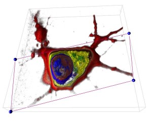
Yann Cotte and Fatih Toy use lasers and 3D photography to visualize living cells.
Two researchers at the École Polytechnique Fédérale de Lausanne (EPFL) have demonstrated their new technique for visualizing living cells in holographic 3D. Using their method cells can be observed over a period of time and the reaction of cells to stimuli can be seen without the use of artificial aids such as contrast dyes or fluorophores, which could distort the interpretation.
The researchers, Yann Cotte and Fatih Toy have combined the use of lasers and 3d camera techniques to create a 3d video. The researchers are working under the direction of Christian Depeuringe, the head of the Microvision and Microdiagnostics Group in EPFL’s School of Engineering. Their work has been published in Nature Photonics. Cotte and Toy use low intensity lasers to avoid exposing the cell to excessive heat. The laser scans the cell sample from various angles and at the same time a digital camera captures multiple shots creating what is a 4D image x,y,z + time. The images can be assembled into a 3D image that can be sliced to expose its internal elements such as nucleus, genetic material, and organelles. As an illustration the team have published a video of a growing neuron as part of the Nature Photonics article.
Toy and Cotte have started a company through the Laussanne startup Lyncée Tec, which concentrates on developing instruments for Digital Holographic Microscopy. Their goal is to use their techniques in order to create 3D observations in vivo, so that live tissue does not have to be removed.
Our take
Is that cool or what?